Compact Bone Diagram Microscope - Microscopic Anatomy of a Compact Bone Flashcards | Quizlet : Each group of concentric circles (each tree) makes up the microscopic structural unit of compact bone called an osteon (this is also called a haversian.
byAdmin•
0
Compact Bone Diagram Microscope - Microscopic Anatomy of a Compact Bone Flashcards | Quizlet : Each group of concentric circles (each tree) makes up the microscopic structural unit of compact bone called an osteon (this is also called a haversian.. The transmitted brightfield digital images above were recorded using a qx3 microscope that was modified for auxiliary illumination. The ground substance of bone is arranged in concentrated layers (lamellae) round the small canals which run parallel to the long axis (shaft) of the bone. Unlike the ground bone specimens, the specimens of two small bones on the following slides were decalcified chemically and then mounted and sectioned with a microtome. Under the microscope dense, compact bone shows a definite and a characteristic pattern of arrangement. Best worksheet compact bone microscope slide labeled free download file 624 diagram of compact bone new jpg wikimedia commons.
A cross section of decalcified compact bone is examined under brightfield illumination with the intel qx3 microscope. Proper use of a microscope. Compact bone is very dense and hard on the outside, and makes up most of the bones in the arms and legs. Having been constructed in the 16th century, microscopes have revolutionalized science with their ability to magnify small objects such as microbial cells, producing images with definitive structures that are. It can be remodeled all throughout life identify the circular vessels in the middle of bone running circumferentially around the vessels.
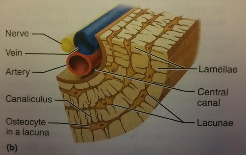
Activity 4: Examining the Microscopic Structure of Compact ... from www.easynotecards.com It's easy to look at these and think of bones as dry, dead sticks in your body, but this couldn't be further from the truth. Each central canal contains blood vessels and nerve fibers surrounded by loose connective tissue. A structural unit of compact bone consisting of a central canal surrounded by concentric cylindrical l. Microscopic osteology and bone formation. The structural units are osteons, which are elongated cylinders acting as. Download de stockfoto histology of human compact bone tissue under microscope view for education en ontdek vergelijkbare foto's op adobe stock. It can be remodeled all throughout life identify the circular vessels in the middle of bone running circumferentially around the vessels. Microscope slide showing a cross section of mammalian compact bone.
Each group of concentric circles (each tree) makes up the microscopic structural unit of compact bone called an osteon (this is also called a haversian.
The transmitted brightfield digital images above were recorded using a qx3 microscope that was modified for auxiliary illumination. Download de stockfoto histology of human compact bone tissue under microscope view for education en ontdek vergelijkbare foto's op adobe stock. The structural units are osteons, which are elongated cylinders acting as. All of our products are students can easily learn the structure of dry, compact bone using this prepared microscope slide. Proper use of a microscope. Compact bone is very dense and hard on the outside, and makes up most of the bones in the arms and legs. Unlike the ground bone specimens, the specimens of two small bones on the following slides were decalcified chemically and then mounted and sectioned with a microtome. Blue histology skeletal tissues bone. When a person is jumping from a certain height or falling by side then a huge amount of imp. Decalcified compact bone at 60x magnification. Static and transient stress long bone stress analysis is very much difficult as for its complex structure (figure 1& 2). Microscopic anatomy of compact bone. Histology of human kidney under microscope view for education hi.
This shows the architecture of compact bone which is designed to nourish and regulate osteocytes and bone matrix. 3 mature bone cells, osteocytes, are found in tiny cavities within the matrix called lacunae. Best worksheet compact bone microscope slide labeled free download file 624 diagram of compact bone new jpg wikimedia commons. Label femur diagram handout • review the following terms: Compact bone is very dense and hard on the outside, and makes up most of the bones in the arms and legs.
Compact Bone Skeletal Sys. Photomicrograph X100 ... from i.pinimg.com Magnification by a microscope is the product of the individual magnifying ability of the oculars and the objectives. If you look at compact bone under the microscope, you will observe a highly organized arrangement of concentric circles that look like tree trunks. The remaining material is mostly most bones contain both compact and spongy bone. A cross section of decalcified compact bone is examined under brightfield illumination with the intel qx3 microscope. Each central canal contains blood vessels and nerve fibers surrounded by loose connective tissue. For example if the ocular is 10x, and objective is 40x, the specimen is magnified 400 times. Unlike the ground bone specimens, the specimens of two small bones on the following slides were decalcified chemically and then mounted and sectioned with a microtome. Trouvez des images de stock de compact bone micrograph light microscopy bone en hd et des millions d'autres photos, illustrations et images vectorielles de stock libres de droits dans la collection shutterstock.
Label femur diagram handout • review the following terms:
Dictionary normal parathyroid gland the human. Unlike the ground bone specimens, the specimens of two small bones on the following slides were decalcified chemically and then mounted and sectioned with a microtome. Label femur diagram handout • review the following terms: Download de stockfoto histology of human compact bone tissue under microscope view for education en ontdek vergelijkbare foto's op adobe stock. When a person is jumping from a certain height or falling by side then a huge amount of imp. Histology of human compact bone tissue under microscope view for education, muscle bone connection and connective tissue. Download scientific diagram | 2: The compound microscope is more complicated than just a microscope with more than one lens. It's easy to look at these and think of bones as dry, dead sticks in your body, but this couldn't be further from the truth. Histology of human kidney under microscope view for education hi. Microscope slide showing a cross section of mammalian compact bone. Blue histology skeletal tissues bone. Microscopic anatomy of compact bone.
Microscopic osteology and bone formation. All of our products are students can easily learn the structure of dry, compact bone using this prepared microscope slide. Overview of microscope and diagram. Label femur diagram handout • review the following terms: There are two basic structural types of bone in mammals, compact and spongy.
6 Osteocytes within compact bone | Download Scientific Diagram from www.researchgate.net Microscopic osteology and bone formation. Microscopic anatomy of compact bone. 3 mature bone cells, osteocytes, are found in tiny cavities within the matrix called lacunae. Trouvez des images de stock de compact bone micrograph light microscopy bone en hd et des millions d'autres photos, illustrations et images vectorielles de stock libres de droits dans la collection shutterstock. A structural unit of compact bone consisting of a central canal surrounded by concentric cylindrical l. Static and transient stress long bone stress analysis is very much difficult as for its complex structure (figure 1& 2). Microscopic structure of compact bone. Two structural arrangements of bone tissue are seen:
Dictionary normal parathyroid gland the human.
Two structural arrangements of bone tissue are seen: Explore microscope parts and functions. It can be remodeled all throughout life identify the circular vessels in the middle of bone running circumferentially around the vessels. 2 compact bone we know that compact bone is very dense it is also very complex when viewed under a microscope. There is a printable worksheet available for download here so you can take the quiz with pen and paper. Microscope slide showing a cross section of mammalian compact bone. The structural units are osteons, which are elongated cylinders acting as. This shows the architecture of compact bone which is designed to nourish and regulate osteocytes and bone matrix. Static and transient stress long bone stress analysis is very much difficult as for its complex structure (figure 1& 2). Compact bone is very dense and hard on the outside, and makes up most of the bones in the arms and legs. Proper use of a microscope. Having been constructed in the 16th century, microscopes have revolutionalized science with their ability to magnify small objects such as microbial cells, producing images with definitive structures that are. When a person is jumping from a certain height or falling by side then a huge amount of imp.
If you look at compact bone under the microscope, you will observe a highly organized arrangement of concentric circles that look like tree trunks compact bone diagram. Unlike the ground bone specimens, the specimens of two small bones on the following slides were decalcified chemically and then mounted and sectioned with a microtome.